


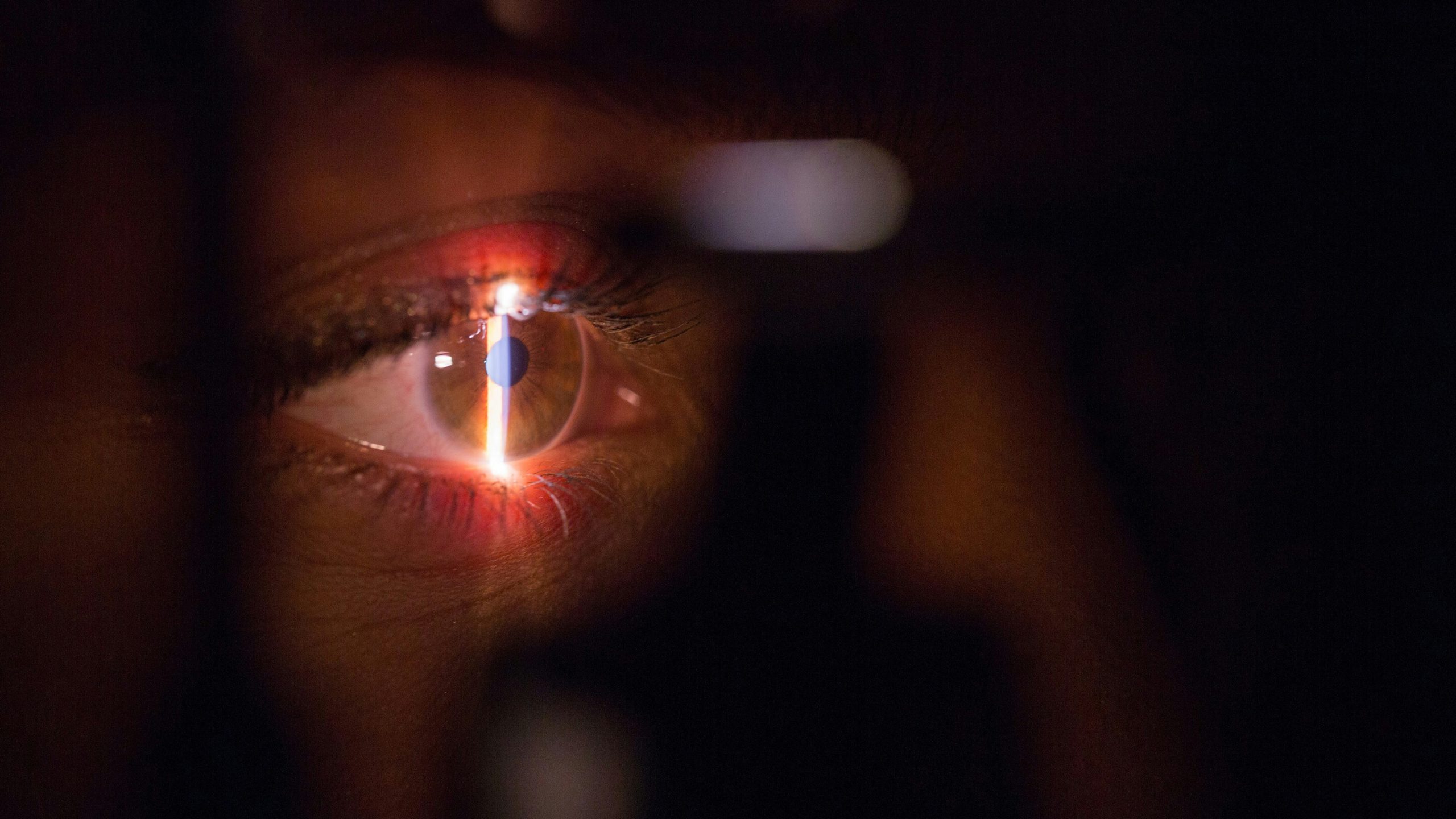
When a person requires cataract surgery or suffers an eye injury, it is common practice to implant an artificial intraocular lens to restore vision. However, in certain patients, the natural structure of the eye that is supposed to hold the lens in place – known as the ocular capsule – is damaged or missing. In these complex cases, ophthalmic surgeons must resort to so-called «secondary fixation techniques», whereby the lens is secured to other nearby structures to keep it stable.
To date, two of the most widely used and advanced procedures for this purpose are fixation to the back of the iris using the «iris claw» (retropupillary) method, and fixation to the white part of the eye, or sclera, using the double-needle suture technique. Although both techniques are routinely used worldwide, scientific uncertainty remained as to which of them ensures better anatomical alignment and greater stability within the eye when the patient moves their gaze.
To answer this question, an international team of researchers – including Pablo Artal, an expert from the Rafael del Pino Foundation’s Chair in Science and Society – has carried out a comparative observational study that provides a pioneering analysis of the position and mechanical behaviour of both lenses in vivo.
The study thoroughly examined 72 eyes from 72 patients, of whom 31 had undergone surgery using the retropupillary iris claw technique and 41 had undergone scleral fixation with clamps. To assess the parameters with micrometre-level precision without invading the participants’ eyes, the team used swept-source anterior segment optical coherence tomography (SS-OCT) and a Purkinje imaging device during rapid eye movements of 9 degrees.
The findings, published in the international scientific journal Journal of Cataract and Refractive Surgery, provide essential data for surgical practice. With regard to the central alignment of the lens with the visual axis, both techniques demonstrated similar levels of accuracy.
In the analysis of lens tilt within the eye, the iris-clamp technique demonstrated a clear physical advantage, recording a lower mean tilt compared with scleral fixation, with a statistically significant difference.
The most striking difference was observed when studying the dynamic behaviour of the lens during rapid eye movements. Lenses attached to the iris exhibited greater dynamic instability or «wobble», characterised by larger oscillation amplitudes following movement, a lower damping ratio and a longer time taken to become completely stationary.
Despite these mechanical differences and the fact that the distance between the lens and the iris was shorter in the iris-clamp group, both techniques proved to be equally effective and safe. The resulting visual acuity, refractive outcomes and complication rates showed no significant differences between the two groups of patients.
The main contribution of this study is to provide the medical community with accurate information on the physical nuances of each technique. Whilst the iris claw results in the lens being less tilted, scleral fixation offers greater resistance to internal oscillations caused by everyday eye movements.